how ear surgeries
are done
The external ear surgery can take as short as 30 minutes to over 12 hours, depending on what ear deformity you have. Sometimes even if one’s ear look the same on the outside, to shape the ear right, a different surgery should be done for the patient. It is important to have an understanding about the difference of the ear structure you are dealing with.
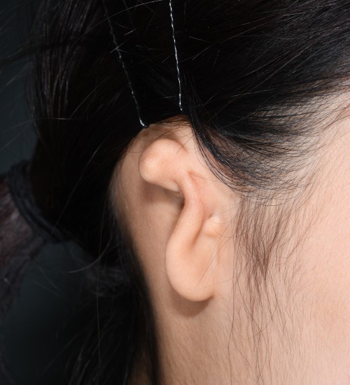
Microtia
People with Microtia have ears that are not fully developed. While the exact cause of this condition remains unclear, it occurs in approximately one in every 8,000 to 10,000 babies.
At our center, we perform Microtia reconstruction for individuals aged 12 and above. We have found that performing this surgery before the age of 12 often leads to less satisfactory results, which is why we do not recommend early reconstruction.
The total reconstruction of Microtia at our center is carried out in three stages, with a six-month interval between each stage. We advocate the use of rib cartilage for the ear framework, as opposed to Medpor. Rib cartilage is a safer and more durable option for reconstructing the ear.
Although some centers use a Medpor framework for ear reconstruction, which may be simpler initially, it can lead to complications over time, often within 5 to 10 years. We have treated several patients who initially had Medpor implants elsewhere and later came to our center for secondary surgery.
related publications
(Dr. park)
- Total Rebuilding of the Ear after Unsatisfactory Initial Microtia Reconstruction: 30-Year Experience using Autogenous Costal Cartilage Framework J Plast. Reconstr. Aesthet Surg. 2023;86:174-182
- Microtia Reconstruction in Hemifacial Microsomia Patients: Three Framework Coverage Techniques., Plast. Reconstr. Surg. 2018 Dec;142(6):1558-1570.
- Reconstruction of Microtias with Constricted Ear Features: A 22-Year Experience., Plast. Reconstr. Surg., 2018 Mar;141(3):713-724.
- Discussion: A Novel Method of Naturally Contouring the Reconstructed Ear: Modified Antihelix Complex Affixed to a Grooved Base Frame., Plast. Reconstr. Surg. 2014 May;133(5):1175-7.
- Discussion: Single-Stage Autologous Ear Reconstruction for Microtia., Plast. Reconstr. Surg. 2014 Mar;133(3):663-5.
- An Algorithm and Aesthetic Outcomes for a Coverage Method for Large- to Medium-Remnant Microtia: I. Coverage in the One-Stage Erect Position., Plast. Reconstr. Surg. 2012 May;129(5):803e-13e.
- Balanced Auricular Reconstruction in Dystopic Microtia with the Presence of the External Auditory Canal., Plast. Reconstr. Surg. 2002 Apr 15;109(5):1489-500.
- An Analysis of 123 Temporoparietal Fascial Flaps: Anatomic and Clinical Considerations in Total Auricular Reconstruction., Plast. Reconstr. Surg. 1999 Oct;104(5):1295-306.
- Subfascial Expansion and Expanded Two-Flap Method for Microtia Reconstruction Plast. Reconstr. Surg. 2000 Dec;106(7):1473-87.
- An Analysis of 123 Temporoparietal Fascial Flaps: Anatomic and Clinical Considerations in Total Auricular Reconstruction., Plast. Reconstr. Surg. 1999 Oct;104(5):1295-306.
- A Single-Stage Two-Flap Method of Total Ear Reconstruction., Plast. Reconstr. Surg. 1991 Sep;88(3):404-12.
Prominent Ears
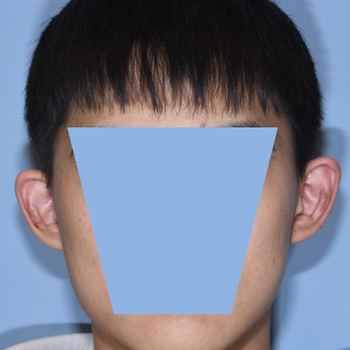
Prominent ears , also known as protruding or ‘bat’ ears, can be a source of stress for individuals, especially after school age when they become more aware of their ears’ distinct appearance.
At our center, correction for prominent ears (Otoplasty) is performed on individuals aged 10 and older, as ear surgery is safer once the ears have stopped growing significantly, which typically occurs by the age of 10. Prominent ears can be corrected using various surgical techniques, including incisionless otoplasty, cartilage scoring, suture techniques, and cartilage excision. While each method has its advantages, they may result in issues such as relapse of ear protrusion, unnatural contours, or sharp edges. Preferences for the degree of ear protrusion vary widely based on cultural or individual differences. Our ear reshaping center strives to create custom-shaped ears that align with the patient’s desire for a natural appearance, minimizing the risk of relapse, through our unique surgical method.”
related publications
(Dr. park)
- Antihelical Shaping of Prominent Ears Using Conchal Cartilage-Grafting Adhesion., Laryngoscope 2012 Jun;122(6):1238-45.
- An Update on Auricular Reconstruction: Three Major Auricular Malformations of Microtia, Prominent Ear and Cryptotia., Curr. Opin. Otolaryngol. Head Neck Surg. 2010 Dec;18(6):544-9.
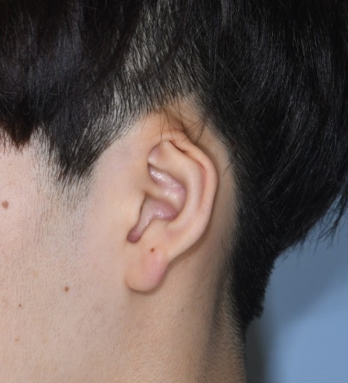
Cryptotia
Cryptotia is a condition where the upper part of the ear cartilage is hidden under the scalp skin. People with this type of ear often struggle to wear masks or glasses, as they lack the normal groove at the upper part of the ear, which typically aids in securing these items. All patients with Cryptotia experience a constriction of the upper ear cartilage, resulting in a deformation that differs from the shape of a normal ear. Additionally, each Cryptotia patient exhibits a unique variation of cartilage constriction. Therefore, the correction of Cryptotia involves not only creating a groove in the upper part of the ear but also requires customized cartilage correction surgery, tailored to each patient.
Correction for Cryptotia is performed on individuals aged 10 and older, as ear surgery is safer once the ears have largely stopped growing, typically by the age of 10. Dr. Park, director of our center center has published a research article on Cryptotia, also referred to as ‘Upper Auricular Adhesion Malformation’. He performs surgery on patients with Cryptotia, who present in various forms, using his developed customized surgical method, ensuring satisfaction for all patients who undergo the procedure.
related publications
(Dr. park)
- Upper Auricular Adhesion Malformation: Definition, Classification, and Treatment., Plast. Reconstr. Surg. 2009 Apr;123(4):1302-12.
- Correction of Cryptotia Using an External Stretching Device., Annals of Plastic Surgery. 2002 May;48(5):534-8.
Constricted Ears
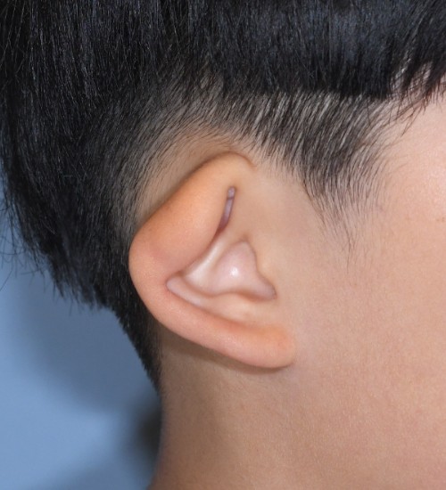
Constricted ears , also known as lop ears or cup ears, present in various forms. While some individuals have normal-sized ears with a constricted appearance, others may have missing parts along with a constricted shape. The severity of a constricted ear can vary depending on the tension in the deformed cartilage.
The surgical approach for constricted ears must be carefully chosen based on an accurate classification of the condition. For constricted ears that are normal in size but simply appear crushed, correction is possible using splints immediately after birth. However, if this initial period is missed, surgery, often complex, becomes necessary for any form of constricted ear.
Dr. Park, director of our center, has published research articles on Constricted Ear, including ‘Classification and Algorithmic Management of Constricted Ear: A 22-Year Experience’ and ‘Reconstruction of Microtia with Constricted Features: A 22-Year Experience’. He performs surgeries on patients with Constricted Ear, who present in various forms, using his developed customized surgical methods, ensuring satisfaction for all patients who undergo the procedure.
Correction for Constricted Ear is performed on individuals aged 10 and older, as ear surgery is safer once the ears have largely stopped growing, which typically happens by the age of 10.
related publications
(Dr. park)
- Reconstruction of Microtias with Constricted Ear Features: A 22-Year Experience Plast. Reconstr. Surg. 2018 Mar;141(3):713-724
- Classification and Algorithmic Management of Constricted Ears: A 22-Year Experience., Plast. Reconstr. Surg. 2016 May;137(5):1523-38.
- A New Corrective Method for the Tanzer's Group IIB Constricted Ear: Helical Expansion Using a Free-Floating Costal Cartilage., Plast. Reconstr. Surg. 2009 Apr;123(4):1209-19.
- The Tumbling Concha-Cartilage Flap for Corretion of Lop Ear., Plast. Reconstr. Surg. 2000 Aug;106(2):259-65.
Earlobe Surgery
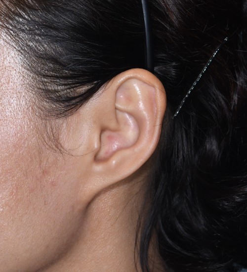
Earlobe surgery addresses a variety of conditions. The most common is the repair of torn earlobes, often resulting from wearing earrings or having earlobe piercings. As specialists in ear surgery, we encounter other conditions as well, including congenital or aesthetic concerns.
One such condition is the congenital cleft earlobe, characterized by smaller, split earlobes. Some centers attempt correction by merely suturing the cleft, but this approach can result in an undersized and rotated earlobe. To appropriately enlarge the earlobe, matching its opposite counterpart, we utilize surrounding tissues in a delicate and complex surgical procedure.
Another condition we address is “pixie ears,” a term for earlobes that appear pulled or elongated. This condition can be congenital, but it is often seen in patients who have undergone facelift surgery. Our approach aims to restore a rounder, larger appearance to the earlobes, adding volume to rejuvenate their look.
related publications
(Dr. park)
- A New Method for the Correction of Small Pixie Earlobe Deformities., Ann. Plast. Surg. 2007 Sep;59(3):273-6.
- Newly Defined Conchal Arterial Floor Flap for Auricular Reconstruction., Plast. Reconstr. Surg. 2002 Jul;110(1):47-53.
- Anatomy and Embryology of the External Ear and its Clinical Correlation., Clin. Plast. Surg. 2002 Apr;29(2):155-74.
- Lower Auricular Malformations : Their Representation, Correction, and Embryologic Correlation., Plast. Reconstr. Surg. 1999 Jul;104(1):29-40.
- Button-Down Procedure for Correction of Cleft Ear Lobe Malformation., Plast. Reconstr. Surg. 1997 Apr;99(5):1429-32.
TRAGUS DEFORMITY
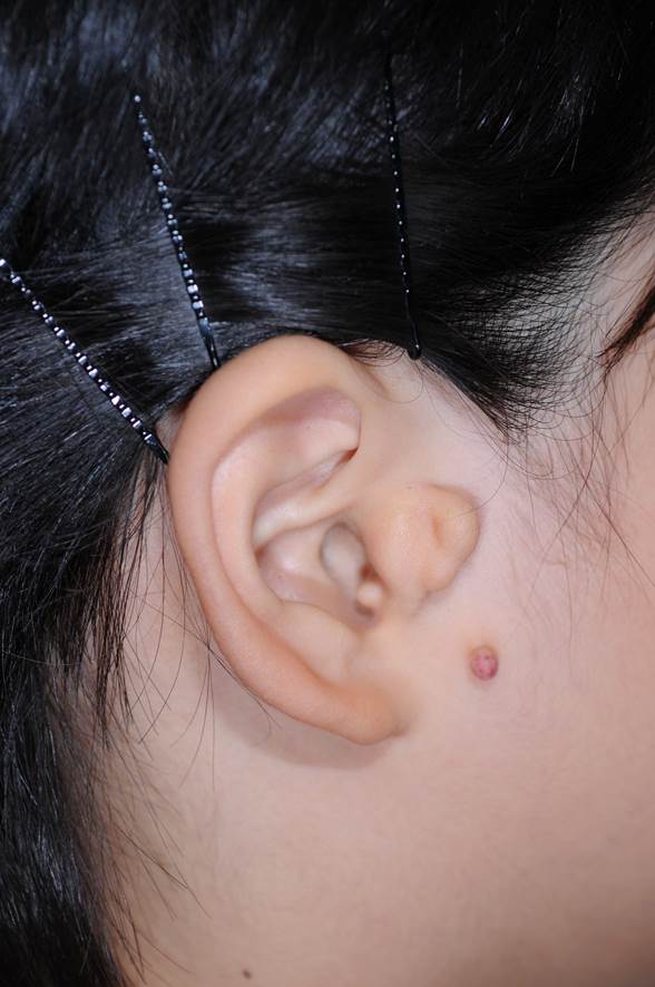
The tragus is a small part of the ear located right in front of the ear canal. It’s a key part of the ear’s appearance.
In East Asia, it’s not rare to see people born with different shapes or sizes of the tragus, often with extra lumps of cartilage near it.
These differences can vary a lot. In severe cases, the ear canal can be fully visible from the side, which might concern some people about their looks. When correcting this, it’s important to make sure the ear canal is not completely visible.
Dr. Park has created a new method to fix this using a special technique that folds and secures the cartilage. This technique has been written about in a medical journal.
related publications
(Dr. park)
- Reconstruction of Congenital Tragal Malformations Accompanied by Dystopic Cartilage Growth (Accessory Tragus) Plast. Reconstr. Surg. 135(6):1681-1692, June 2015.
AURICULAR MARGINAL DEFORMITY
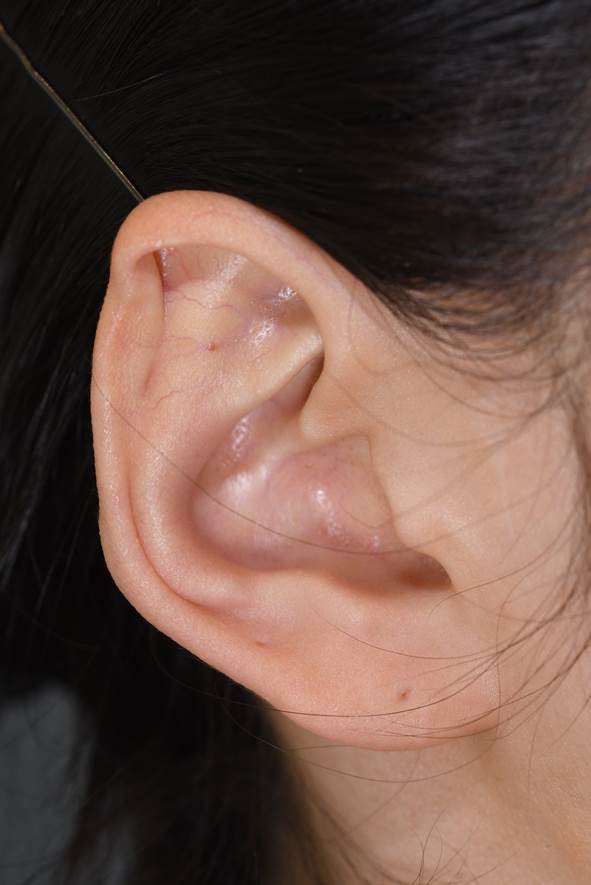
Some people visit the hospital because they are not satisfied with the shape of the edges of their ears, known as the auricular margin or the rim of the ear. This can often be due to the way they were born. There are a few common types of these ear shapes:
1. Helical Adhesion: This is where the helix, the outer rim of the ear, is too close to the front.
2. Shell Ear: In this case, the helix is thinner than usual.
3. Stahl’s Ear: This is when the ear rim is misshapen or bent.
The treatment for these conditions varies depending on how severe the deformity is. For mild cases, we can correct it by adjusting the surrounding skin. However, more severe cases might require reshaping the ear cartilage. Each surgery is unique and depends on the surgeon’s skill and creativity.
Another reason for missing parts of the ear rim can be due to accidents. These cases usually need more complex surgeries than those for birth defects.
Other Ear Deformities
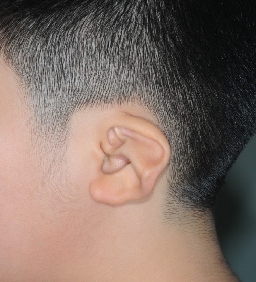
Some ear deformities are not very common, and there’s not much information about them or how to treat them in medical books. Dr. Park, the head of our Seoul Ear Center, has developed new methods to treat these rare ear deformities based on his 35 years of experience treating many patients. Some of the unique ear deformities that Dr. Park has successfully treated include:
1. Question Mark Ear Deformity
2. Concha Striae
3. Spoon Ear Deformity
4. Others
It can be challenging even for a plastic surgeon to diagnose these uncommon ear deformities if they don’t specialize in ear surgery. So, if you have any unusual ear shape or concern that you want to discuss, please consult us.
related publications
(Dr. park)
- Reconstruction of Congenital Tragal Malformations Accompanied by Dystopic Cartilage Growth(Accessory Tragus)., Plast. Reconstr. Surg. 2015 Jun;135(6):1681-91.
- A Single-Stage Two-Flap Method for Reconstruction of Partial Auricular Defect., Plast. Reconstr. Surg. 1998 Sep;102(4):1175-81.
- Correction of the Unilateral Question Mark Ear., Plast. Reconstr. Surg. 1998 May;101(6):1620-3.
- An Improved Burying Method for Salvaging an Amputated Auricular Cartilage., Plast. Reconstr. Surg. 1995 Jul;96(1):207-10.

