
The Seoul Ear Surgery Center is the single most experienced center for ear anomalies throughout Korea. We have more than 2000 microtia patients reconstructed in our center for over 30 years. The senior surgeon, Chul Park also has shared is vast experience to the “American Society of Plastic Surgeons” with numberous publications. (you can also search on google as “chul park md plastic and reconstructive surgery” to see his work)
For typical lobule type microtia, reconstruction in our center can be done at any age after 12. When the reconstruction is done before such age, result would be less satisfactory (which we do not recommend). Total reconstruction of microtia is done in 3 stages with 6 months interval. We recommend using costal cartilage for the framework of ear (not Medpor), which is the safest and durable way of reconstructing the ear. Some other centers are using “Medpore” framework for the ear, which is easier, but it is likely to make problems eventually (over 5 or 10 years later). Patients who have problems with Medpore come to our center later on, to get a secondary surgery.
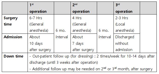
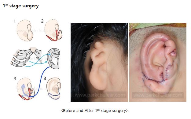
The 1st stage of reconstruction is done in general anesthesia. We harvest your own rib cartilages from the rib cage, and carve to make an ear frame which resembles the opposite ear. Then the carved ear frame is inserted under the skin where the ear should be located. After the 1st stage, as ear shape is buried under the skin, you cannot see the exact shape of the reconstructed ear nor the ear frame. The surgery takes about 6-7 hours, and you need to stay in the hospital for 10 days after surgery.
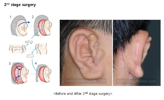
The 2nd stage is also done in general anesthesia. On this stage, we are elevating backside of the framework in a protruded state so that we can see your both ear from the front view. Back side of the ear framework is then covered with additional tissue (fascia) and skin. The surgery takes about 4 hours, and you need to stay in the hospital for 1 week after surgery.
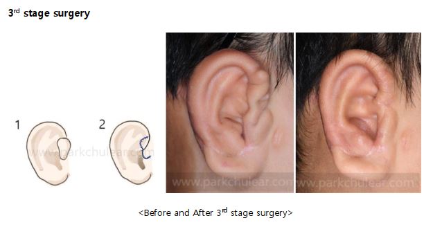
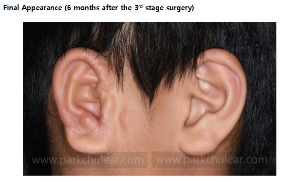
The 3rd stage which is the last stage of the surgery, is done by local anesthesia. We remove the remaining skin from the reconstructed ear to make the shape more close to the opposite ear. We are not making an earhole, but it will look like as if you have an ear hole of your own. (We do not recommend making a hole in the area since we have seen too many patients suffering from problems after the earhole surgery) The surgery takes about 2~3 hours, and you will be discharged on the same day after the surgery.